Wednesday, November 4, 2009

Midterm Season!


So, yes, epic fail on my part for keeping up with the labs and pics and little nuggets of interesting info. Ever since leaving the world of muscle and bone the drawings have been less interesting and the knowledge much more factual. Don't get me wrong, it IS really fascinating, it just lacks the that specific artistic. But I haven't been COMPLETELY useless the past few weeks! Reaching for some classical anatomy I went to a figure drawing class on campus last saturday (before going off to lab to study the body's insides) and got a few cool drawings out of it. It was really incredible to draw after learning the muscles since I could see the anatomy in action. Plus, it helped that our model was a super fit, younger, dancer/actor/acrobat (ie. awesome body). For those of you not familiar with figure drawing, YES its entirely in the nude, and YES you get models of all shapes and sizes (one time I had a man who must have been 70 and his size and beard made him disturbingly similar to santa clause--but hey, its art! (right?) )

Tuesday, October 13, 2009

Lab 3: The Humerus is NOT a funny bone....or maybe just a little....


SO. with the busy life still unrelenting, I have decided to post pics before its TOO late from the third lab - explorations of the arm bones, and their muscles.

A couple comments...

Rotator cuffs are awesome! Who would have thought a succession of so many muscles allowed the arm's wide array of movement

The humerus is not a funny bone!
although its name is humerus...
For the 99.9% of people who have ever awkwardly bumped their elbow to have a "funny tingle"
(more accurately it's NOT funny it's painful! or maybe my arm is unusual...) has not actually bumped the "elbow" but the medial (inside) epicondyle (bump) of the humerus (upper arm bone). By bumbing this bony projection, the Ulnar nerve gets pinched and sends a strange tingling sensation through the arm. I guess for kids it's funny, and at least there is some truth behind the colloquial name for the bone!

BUT don't get confused! the "bony part of the elbow" that you rest on a table or scrape when you fall is not the humerus, but part of the Ulna (a forearm bone). (the fancy name for this projection is Olecranon process)


Wednesday, October 7, 2009

It's Been a LONG Time...

...since I last wrote. Lab got so busy and crazy that as I tried to stuff the last little pieces of knowledge into my brain the blog was left by the wayside. A quick overview of what was covered since I last wrote: Lab 3: The upper extremities (the arms!), Lab 4: The lower extremities (and legs!), and The midterm practicum. All three things huge events worthy of their own postings... I might have to give them due justice.

For now, a few reflections.
Into the first week of october and it seems that the course has covered everything I was artistically interested in. Although the muscles were a pain to memorize, it actually got fun tho think it all out:

"so if the Gastrocnemius (calf muscle) attaches to the back of the tibia"
(feeling behind my knee)
"and the calcaneous bone of the foot via the Achilles tendon"
(and feeling my heel)
"then if I contract it, my foot dorsiflexes!"
(and voila, pointed feet!)

muscle by muscle I would study. Rotating arms, twisting legs, moving my wrists this way and that to learn by doing and feeling the action.

Now that I KNOW what each muscle does (at least I hope I do!) I am even more excited to get back to figure drawing and have that realization
"ahh! flattening the hands contracts the extensor digitorum causing the slightest bump in the arm!" and see it on the model.

But now we move on, a new lab bright and early tomorrow... so I bid farewell to the beautiful anatomy of musculature and dive deeper into the cadaver's systems. Digestion. Excretion. Yay? While curious, they offer less obvious artistic applications. Hopefully this lab will produce some creative rederings of kidney lobes and intestine, yet I'm guessing they will lack the aesthetic beauty of the body's muscles bones and sinews.

I'll have to just get to drawing and see what happens, since formless complexity can also be beautiful (Pollack anyone?)

Thursday, September 17, 2009

DRAWING the anatomy

DRAWING the layers of muscles--one weaving into the other in a seamless beauty; it's a testament to the complexity of what can seem so simple.

I actually got excited when I got to draw the sternocleidomastoid since it is a muscle I have SEEN on models in figure drawing class. It's a muscle that is clearly visible on Michelangelo's exaggerated ignundi and his David and other renaissance depictions of the ideal man. When the head twists in such a way you can see its contraction and tension.


(my own "illegal" pic of Michelangelo's David from this past spring when I was in Florence! Check out the sternocleidomastoid muscle that goes from his ear (mastoid process area) to the top of his breast bone (sternum) incredible.....)

It's one of those muscles that bulges forth from beneath the skin--defining itself against the rest of the ordinary flesh. It is beatiful in form--and it is just as beautiful in function. As the incredibly long name suggests it has many attachment points (sterno = sternum, cleido = clavicle, mastoid = mastoid proceess of temporal bone). And with all these attachments, it can do wonders when it contracts. Just try tilting your head forward, then back, and then from side to side: all of those movements use one or both sternocleidomastoid muscles (with some others) it's versitality is remarkable.

Speaking of drawing.... after lab I was still trying to figure out the deep muscles of the hip that we had "seen" on the cadaver (the UGSI pointed them out but I was clueless!) I decided to draw the muscles and got carried away and voila the leg! We haven't officially learned the muscles bellow the iliacus but it was fun to draw them all out anyway. Legs are so complex to draw that just seeing the muscles helps it make so much sense. Leonardo's sketches incorporate beauty while retaining incredible scientific accuracy. wow.

LAB 2: Rectus abdominus + tendinous intersections + crunches = 6-Pack

Rectus Abdominus is the official name of the colloquial "abs" or for those lucky enough, "six pack." Unfortunately last Wednesday when I was taking the pre-lab quiz I completely blanked on what muscles are in the abdomen. Sure I could point them out but thanks to a last minute essay I had to write I hardly studied and had no clue for their names! NOW I know, and as long as I'm in lab I'll use the official name instead of my colloquial interpretation.


So, the second lab came and went in a confused array of muscle names and histology slides. Although in this lab we got to work with cadavers, it felt significantly less hands on than the first week. The cadavers were real, but other than that were mostly observed and everything was explained to us by a UGSI (undergraduate graduate student instructor--I know it makes no sense, Berkeley is odd and doesn't want to use the universal term "Teachers Assistant" TA).

Compared to the bones which I got to hold, explore, turn over, and examine in the first lab, the cadaver was so academic and brief. I even attempted to make a drawing of the back's muscles which ended up resembling a piece of wood...

One thing that the cadaver experience DID show was the amazing array of muscles that make up the neck! The thin platysma muscle hides so many criscrossing and varied muscles. Everything from the sternocleidomastoid that turns the head to the geniohyloid that helps with swallowing: it's all there (and on the cadaver it all looks like the same pink-brownish meat). Somehow by the end of all of this I'll be able to point at one of those random muscles and know it's name attachment points and function (hopefully!)

While anatomy can be daunting at times, it can also be incredibly self explanatory. For example, on our quiz (which I ultimately failed...) one question was "What is the function of the erector spinae?" Erector Spinae? Erect spine? Holds the spine erect? Ka-ching! my logic worked! unfortunately not EVERY muscle is so kind, but it was exciting to figure it out after starting without a clue!

And in case you are still reading....
some cool random factoids I encountered in Lab!

Geniohyoid muscle = helps with swallowing but is REALLY hard to see on a cadaver because like the genie in a bottle its hidden!!

ligamentum nuchae = string on the back of the neck that literally holds your head up-- I had no idea that yoga/ballet teachers were referring to an actual string!

linea alba = even basic latin should tell you "white line" and YES another self explanatory term: its literally the white line that runs down the midline of the abomen in line with the belly button. This difining midline of the 6-pack is made up of tendonous material and is actually white. The linea alba always reminds me of Diamonds lecture on the abdomen muscles and c-sections. Apparently originally caesareans cut along this linea alba since it is a natural "opening point" into the abdomen. However, this method was more prone to complications in healing so the surgery switched to a lateral (horizontal cut) just below the bikini line. Last year Prof. Diamond joked how the ladies didn't like having a scar, but didn't mind having so many torn muscle fibers (since a horizontal cut goes through a lot more muscle!) Apparently the lateral cut goes through more muscle for mainly cosmetic reasons. How random science is some times! Sorry for the digression....

One thing I did learn is that I take much more pleasure in simply drawing the muscle groups and bones than knowing every little name for every bulge tubercle or fossa. However if I want to speak coherently about anatomy I'll have to learn the language. At the very least I will be prepared the next time I go to the doctor and I can tell them the exact bone or muscle that hurts!

Lab 2. check. a little trickier than the first, a lot of material, but I am sure slowly and surely this will all start to catch on (as long as I don't start reciting anatomical knowledge in my sleep!)

Oh! and if by now anyone is still reading feel free to check out the pics in better resolution--just click on them. I was too lazy to crop out the uninteresting stuff (and I never know what is the most interesting!)

ciao ciao tutti!

Reflection on Working with the Cadaver!

**Technically this is not my next post! Rather, it is an essay I wrote FOR my class so I apologize for the academic style. I was responding to the prompt below,

"We would like you to reflect on your first encounter with the cadaver. . Whether it made you sad, excited, or frightened, be genuine in your responses as there is no wrong answer."

feel free to read!

and an update from the lab is coming next :)

Although it is tempting to claim that encountering the cadaver made me contemplate my mortality or scared me in some way I cannot—unfortunately the encounter was so built up and anticipated that when it actually happened it was almost mundane. Honestly I was expecting a flood of emotions: questioning the strangeness of studying the muscles and sinews of a form that not too long ago had a life, a story, a family, and a name. But no, I was as cool as a cat (assuming cats are cool) and didn’t even mind touching the cadaver to make sure I had noted the right muscle group. The cadaver seemed more like a plastic model than anything else, and the experience was unlike what I had expected, almost underwhelming. And by underwhelming I do not mean to suggest that it was not intellectually stimulating, since seeing all the muscles in their natural form (not color coded and text book perfect) was quite an experience. But it was not disturbing as I expected it to be.

After thinking about it, I understand a little better why emotionally I was fine with studying the cadaver. In high school I volunteered at the Science Center in LA and worked in the Body Worlds exhibit where real cadavers are preserved by plastination and on display in various poses or in certain slices to help demonstrate some muscle group or body system. By being in the exhibit so frequently I grew comfortable with seeing entire cadavers—face, hands feet—nothing covered up, and the entire thing stained in colors representative of living tissue (reds etc). Because of this I knew I would be more prepared to see the cadaver, but I was not sure how hands on the anatomy 131 lab would be. However other than the slight touch of a muscle and the strong smell of the preserving solution, the lab was mostly visual, similar to the exhibit. And the cadavers were so prepared—everything cut and pulled back and extra stuff removed. Honestly, I was more disturbed by the rat dissection in Biology 1A lab where we actively cut open through the rat skin and sought out different organs (while the rat was still a little warm). Comparatively the experience with the cadaver was stale, distant and mostly visual.

As stated, emotionally I was unaffected, but intellectually the cadaver was a wonderful way to study. I was surprised by the layering and compact nature of many of the muscles. I also found it intriguing how area of the body that seems simple on the surface can be so complex underneath. Especially interesting was how flat and undefined many of the muscles were. In light of my observations of the human body in action; whether in athletic events, dance competitions, or in figure drawing classes, I was expecting much more definition for the individual muscles. I even got out my sketchbook and tried to draw directly from the cadaver but it was unfortunately rather boring. The drawing came out very flat and uninteresting. The over exaggerated muscles portrayed in the anatomy coloring book are much more fun to draw (since they are over defined). I’m not sure why there is such a strong discrepancy between muscles of living bodies and those on cadavers other than the obvious: cadavers are carefully preserved and lack the activation, water, and whatever else that makes animated muscles so visible, also I am used to seeing younger bodies while cadavers can be of any age group I'm guessing.

While my reactions were not what I expected I realize that they were my reactions just for the first lab. With time and with subsequent labs I’m sure that my perspective will shift as we explore different areas of the body. Also as I get more comfortable with the set up it will hopefully be more interactive and less observational. The human body is such an incredible thing that to finally study the real thing after so many years of learning through books and pictures it is hard to know how to feel. At the very least I am still excited for what the next lab will bring.

Tuesday, September 8, 2009

Make no bones about it!


Lab 1: check!

NOW I finally feel in tune with this anatomy class. While in lecture I feel like I'm trying to learn by seeing shadows on a screen, the Lab is refreshingly hands on and personal--I felt like Alice stepping through the looking glass* by actually getting to touch, examine, and question my surroundings instead of simply passively accepting the image fed to me in class. *(disclaimer! I have not read Alice and the Looking Glass, although I'm assuming she steps through some sort of mirror into some crazy world if good ole Disney has taught me anything!)

Last Wednesday I walked into lab and, diverting my eyes away from the back of the room where two stretchers covered by sheets waited (cadavers that we will be investigating this week!) I walked straight to my lab bench and sat amidst boxes and boxes of bones. Some were stringed together in order, other were jumbled up randomly, a few were color coded (most were not) and all of them were real bone-a-fide human bones! (sorry for the cheesy pun :)

Looking into a box of the crazy beige colored shapes, I couldn't help but be reminded of the first time I saw written Greek; sure I knew it was a language, I knew the letters pieced together to words, and I knew the words could be read to make sense, but I still had no idea how to understand any of it! It just looked like jumbled symbols. Same with these bones: "yes, that is definitely a rib, and this a skull, but what are all of THOSE? vertebrae!? so THAT's what they look like! What's that shell thing doing here?" But just as the vague similarity between some Greek letters and Latin letters allows basic words like the names of gods or places to be legible, the analagous shape and structure of the many bones to the assembled skeleton started to make the seemingly random vertebrae pieces much less random.
Par example, I've got no idea who the gods are on the right but I'm pretty sure that the one on the left is labeled "Dionysus"


At one point I picked up two vertebrae to see how these crazy shapes could fit together and voila! like two puzzle pieces they slid one into the other in beautiful alignment (luckily I had chosen two thoracic vertebrae, so this actually worked!). Yes, it was starting to make sense, slowly the foreign was becoming familiar. As I worked through the lab book and looked for different characteristics of the bones, I started to see the subtleties while learning the anatomical language. Among other discoveries, apparently foramen is the cool way to say "hole"
similarly...

process
= bulge
fossa = pit (its Latin for trench! thank you wikipedia :)
facet = where another bone will meet/touch
articulate = same as facet (except it will move)?
and then there are the words that thankfully define themselves (angle, groove, arch)

Sorry to bore with the anatomy lingo, it just seemed so cool that I could sound very knowlegable and point out the "vertebral foramen" aka "little holes in the piece of backbone." Although it is probably a good thing the anatomical terminology is there, my discriptions are too long to say over and over again!

So after examining drawing and piecing together the many different bones I soon enough realized that I was running short on time and there was a WHOLE lot more to do. All the microscopes were still sitting with the histology slides (histology = "study of tissues" in Greek! basically microscopic slices of everything from skin to the eyelid of a monkey). It was deja vu all over again except the random vertebral bones were now pink and purple blobs under the microscope. "Ah! there they are! the stratified columnar cells! oh wait, those aren't cells? ahh... I'm looking at the connective tissue apparently, gotcha...." VERY slowly the histology slides started to make sense, although I must admit handling human bones is a little cooler than hunching over a microscope.

So, to recap, a few cool realizations...

Biological Puns!

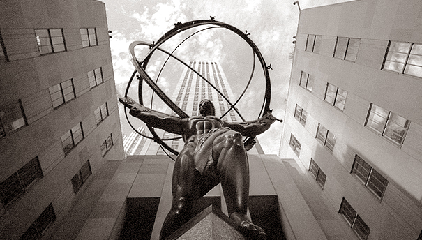
Someone out there in the anatomical world was clever when they named the first two cervical vertebrae the atlas and the axis since the first vertebrae, the Atlas (named after our friend from Greek mythology and the guy who holds the big bronze gyroscope in Rockefeller Center) holds up the "globe" of the head, and the Axis acts as its pivoting point! one thing I'll hopefully remember.


Form and function!

Everything from the "little holes in the vertebrae" to the random bump seems to have a function for some nerve, muscle, or other bone--it's really incredible how the body which seems so random (looking at pieces of skeleton) can make perfect functional sense. Now just to figure it all out.

The skull...

is beautiful! So that's why cheekbones have that shape, why the nose varies, why the brow can create the deep set eyes... Drawing the skull really proves the beauty of the human face. So many bones fused almost seamlessly together into one undulating recognizable form.
At one point I held the skull in my hand to study it and couldn't help but think, "Poor dead jester-guy from Hamlet!" (Since lab, I have since looked it up, poor Yorik would be sad his name wasn't more well remembered :)

The most incredible thing about the skull is that it seems to be one solid form with, okay, the jaw bone is probably seperate, but really how many bones can there be? Granted they are all fused together*, but there's an impressive total of 8 cranial bones and 14 facial bones of every crazy shape and size!

*so apparently the Hyoid bone is NOT fused to the skull, which is why its usually lost, but it is nevertheless the important base for tongue muscles. another anatomical fact you didn't want to know!!!

anyway. enough of my musings. now to do some reading for the NEW lab tomorrow, where we'll aparently dive (not literally....too soon?) into cadaver dissections! Yay!? Nervous and excited?

ciao tutti! until next time!

Friday, August 28, 2009

Here's hoping.....

that my expectations won't be too dashed with the last minute change in my anatomy course. After all my excitement and anticipation I showed up to the first day of lecture this past wednesday, and there was Professor Diamond with her bright white hair, and her infamous floral patterned hatbox. As the lecture hall of some 700 plus VERY loud students settled down she calmly set the big hat box on the podium, opened it up, and pulled out a human brain. While the hall was still buzzing with all the students, she gave her opening lecture on the brain and its incredible capacity to comprehend and imagine despite weighing only 3lbs. The joy of her simple lecture style was short lived however, and she announced that for the FIRST time in 50 years she is NOT teaching the class! Because of torn rotator cuff muscles she cannot write on her big blackboards and will be taking the semester off. (okay, she IS at least 83 years old, I guess she's entitled to take a break). soooo she is handing the class off to the summer session teacher, a very good professor but very different.

Goodbye chalkboards, hello power point--slides that are read over so fast there is no time to write them down and barely enough time to comprehend. We are expected to go back and learn the material and read it on our own time (a very different approach compared to the IN class learning Diamond encouraged). Sure we are asked questions and given about 2.5 seconds to answer before boom! the next slide is up and we are off and moving into new material. I sat through nearly an entire semester of Diamond's anatomy class last fall and the differences are striking (I was not able to ACTUALLY enroll because Berkeley is freakishly impacted, and was perpetually on the waitlist).

Instead of condensing the information into accessible concepts and principles we are given the worlds supply of information and left to pick up all the pieces to make it understandable. Diamond would have the class repeat difficult words, encourage drawing the structures to understand them, and overall ENGAGE each and every student in the 700 person lecture hall so the class never seemed too big. And every Wednesday she would pick four students at random to have lunch with at the faculty club. Learning was about talking, sharing, questioning, and drawing. Sadly, a very different semester lies ahead, one where the learning occurs at home or in the library hunched over a fat textbook instead of in the classroom. Where participation is charted through electronic i-clickers (remote control answering device things) and notetaking and drawing are practically discouraged (since all the slides are available online allowing us to simply listen to lecture). Although I'm really happy that I got the chance to audit Diamond's lectures, it's too ironic that when I finally get the chance to take the class, it's no longer the same. Ahh c'est la vie!

At the very least I'll attempt to be extra dedicated to drawing and journaling and once lab starts next week I'll hopefully have some cool pics to post! I'm pretty sure it will all be skeletal but its exciting nonetheless.

ciao for now!
here's hoping....

Wednesday, August 19, 2009

or the Anatomy of Art?

Ciao tutti!
So with my week of summer upon me I'm feeling ambitious and in need of something else to do. Hence a blog (that I will hopefully keep up!) that will follow my simultaneous interests of science and art. I will finally be taking Anatomy Lecture and Lab (ie. Integrative Bio 131) at Cal this fall, after getting kicked out of the class last fall (the waitlist was too long!) The lecture is taught be the lovable Marion Diamond, who despite her age (83?) insists that students answer questions during lectures and takes 3 students out to lunch every week. She is traditional and uses the big chalkboards--refreshing in an age of powerpoints and digital learning. Although the once-a-week lab is far more intimidating, it does involve analysis of human skeletons and cadaver dissections. As excited as I am to cap off my academic premed track, I'm even more excited to sneak my sketchbook into my lab room and get to drawing. To join the ranks of Michelangelo and Da Vinci and their unsatisfied curiosity for the beauty and complexity of the human body behind the flesh.

So what's the point of this blog?
I'm not sure where it will go, but for now I hope to post musings/reflections on the class and drawings that I make during lab. Hopefully I'll draw some sort of connection between the Art of the Renaissance and the Art of a modern med student, if not, I'll have some cool drawings of muscles and sinews by the end of the semester!

ciao for now!